Calcein AM/PI Double Staining Kit
Calcein AM is the addition of an acetyl methoxy methyl ester (AM) group to Calcein, which increases its hydrophobicity, allowing it to easily penetrate live cell membranes. Calcein AM itself is non-fluorescent. Once inside the cell, it is hydrolyzed by intracellular esterases to produce Calcein, a highly negatively charged, polar molecule that cannot pass through the cell membrane and thus stays inside the cell. Calcein emits strong green fluorescence (Ex/Em = 494 nm/517 nm). Since dead cells lack sufficient esterases, they cannot or only weakly produce Calcein, resulting in strong green fluorescence in live cells and minimal or no staining in dead cells. Dead cells, having compromised membrane permeability, allow Propidium Iodide (PI) to enter and bind specifically to double-stranded DNA, producing intense red fluorescence (Ex/Em = 535 nm/617 nm) to mark dead cells. Thus, the combined use of Calcein AM and PI enables simultaneous dual fluorescence staining of live and dead cells, useful for assessing cell viability and cytotoxicity.
Elabscience® Calcein AM/PI Double Staining Kit can be used to distinguish between dead cells and living cells in mammalian samples with esterase activity.
Features of Elabscience® Calcein AM/PI Double Staining Kit
Wide Range of Applications:
Suitable for a variety of suspension cells and adherent cultured cellsDiversification of Detection:
The results can be detected by flow cytometer and fluorescence microscopyEasy to Operate:
No need to spend time on reagent concentration adjustments, only takes about 15~30 minLow Toxicity:
Doesn't affect cell differentiation and proliferationCost-Effective:
The reagent component is complete, and the buffer contains components that prevent Calcein leakageCalcein AM/PI Double Staining Kit and Related Reagents
| Product Name | Size | Price |
|---|---|---|
| Calcein AM/PI Double Staining Kit | 100/500/2000 Assays | More Details |
| Calcein AM Solution(100 µM) | 100 T/500 T/500 T*10 | More Details |
| PI Solution(750 µM) | 100 T/500 T/500 T*10 | More Details |
| Calcein AM Assay Buffer | 100 mL | More Details |
The Results of Calcein AM/PI Double Staining Kit
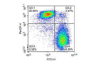
Figure 1. Jurkat cells were placed at 4℃ for 20 days, then stained with Calcein-AM / PI Double Staining Kit and detected by flow cytometer.
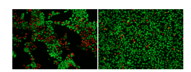
Figure 2. 4T1 cells (left) and Hela cells (right) were treated with 5 μM Camptothecin for 4 h and photographed.

